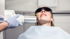
X-rays in dental care
X-rays are used in dental care to support clinical research. The images give the most accurate picture possible of the condition of the teeth.
X-rays in support of dental care
With the help of an X-ray, it is possible for the dentist to detect various infections hidden in the mouth as well as cavities that have penetrated the enamel at an early stage. Early detection of inflammation greatly improves the prognosis of treatment. X-rays are a reliable way to detect other diseases that are hidden in the mouth. With the help of a dental x-ray, the dentist assesses the condition of the jawbone and teeth, among other things.
The dentist will assess the need for an X-ray for each client individually. X-rays can be performed by either a dentist, oral hygienist, dentist, or radiologist.
Different forms of X-ray imaging
Most commonly, single x-rays are taken of the dentistry during dental care. These include periapical and Bitewing images. From the periapical image, the tooth can be inspected from its crown right to the very tip. Periapical imaging is necessary, for example, in connection with root canal treatment. The Bitewing images, on the other hand, show cavities hidden in the gaps between teeth and tartar for example. Bitewing images are often taken during a basic oral examination.
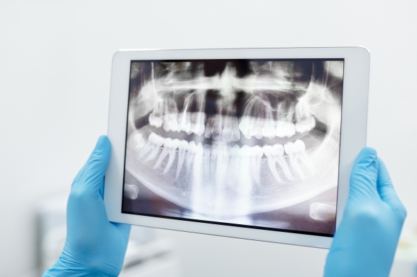
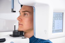
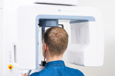
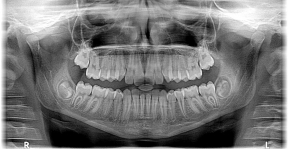
In addition to periapical and Bitewing images, panoramic X-rays can be taken of the dentition. The panoramic image shows the jaws, jaw joints, buccal cavities and the dentition itself.
Panoramic X-ray imaging
Bitewing X-ray imaging
